详情
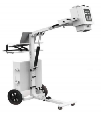
1.Overview SimulatestheentireworkflowofaportableDRsystemformobiledigitalX-rayimagingofthehead,cervicalspine,limbs,chest,abdomen,andotherbodyparts,facilitatingteachingandtrainingtasks. 2.MainComponents: 2.1MobileFrame 2.2ModularHigh-VoltageGeneratorPhantom 2.3Flat-PanelDetectorPhantom 2.4Collimator 2.5DigitalIntelligentImagingPlatformWorkstation 2.6DigitalIntelligentImagingPlatformSoftware 3.KeyTechnicalParameters: ▲3.1MobileFrame:Movableandwheeledforclinicalmobileimaging. 3.1.1Verticaltravelrangeoftelescopicarm:≥1300mm 3.1.2Maximumfocalspotheightfromtheground:≥1800mm 3.1.3RotationrangeofmodularX-raytubeassemblyrelativetothemobilearm:≥±180° 3.1.4RotationrangeofmodularX-raytubeassemblyrelativetoitshorizontalaxis:≥120° 3.2ModularHigh-VoltageGeneratorPhantom 3.2.1Simulatednominalpower:≥5kW 3.2.2Simulatedtubevoltagerange:≥40-120kV 3.2.3Simulatedexposurecurrentrange:≥5mA–100mA 3.2.4Simulatedexposuretimerange:≥0.002s–10s 3.2.5Simulatedcurrent-timeproductrange:≥0.1mAs–250mAs 3.2.6Simulatedexposurecontrolmethod:Handswitchexposure 3.2.7Frameincludesastoragecompartmentfortheflat-paneldetector. ▲3.2.8Softwareconsoleishighlyintegratedwiththehigh-voltagegenerator,allowingexposure parametersettingsviaPC. 3.3Collimator 3.3.1Realmedicalcollimator 3.3.2Lightfield:Continuouslyadjustable 3.4Flat-PanelDetectorPhantom 3.4.1Detectorsize:≥17×17inches 3.4.2Quantity:≥1unit 3.4.3Simulatedpixelsize:≤160μm 3.4.4Simulatedpixelmatrix:≥2880×2880 3.4.5Simulatedgrayscaledetection(A/Dbitdepth):≥16bit 3.4.6Finalimagegenerationtime:≤3s